Advanced Microscopic Neuroimaging
ZEISS Elyra 7 Lattice SIM²
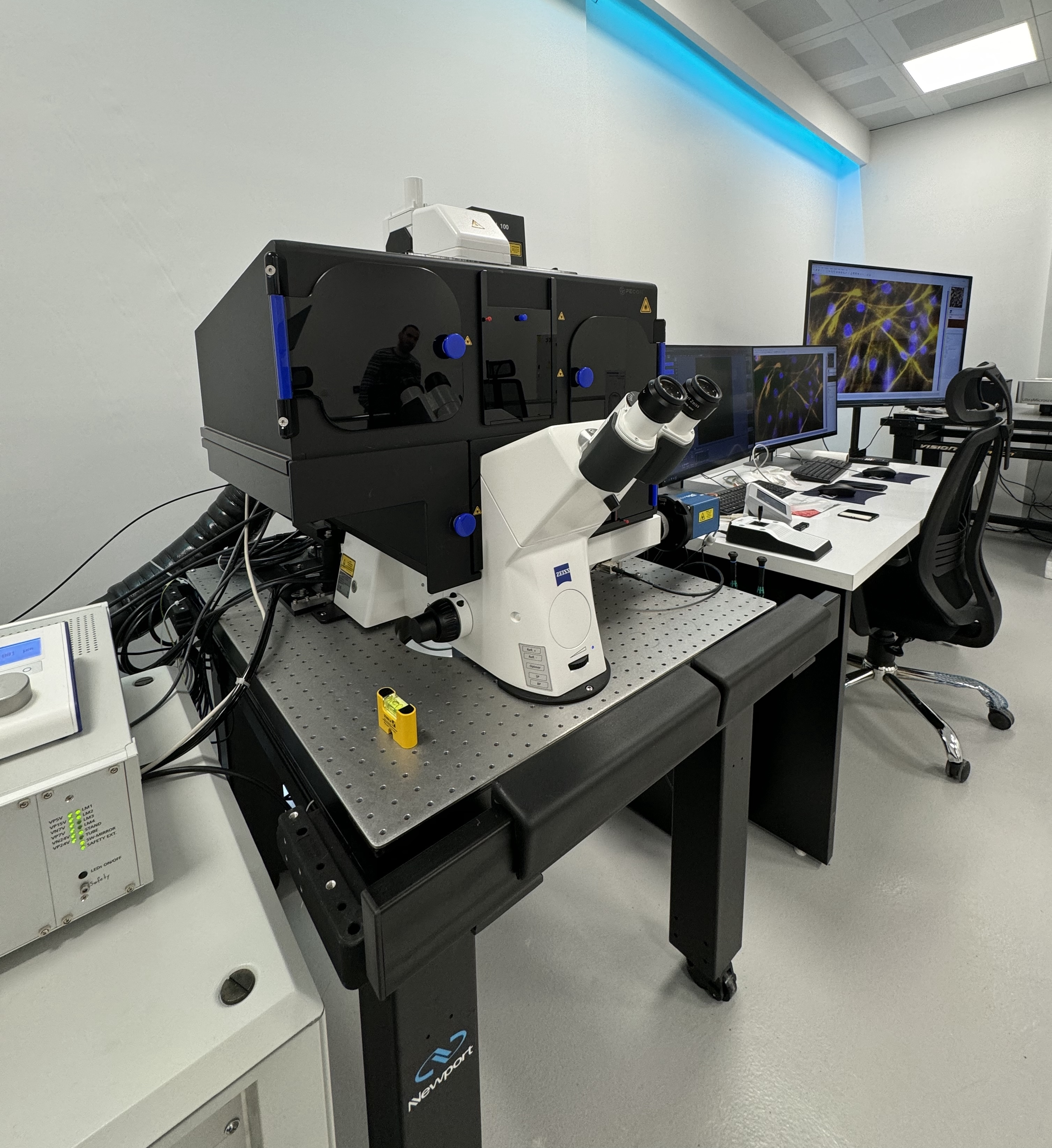
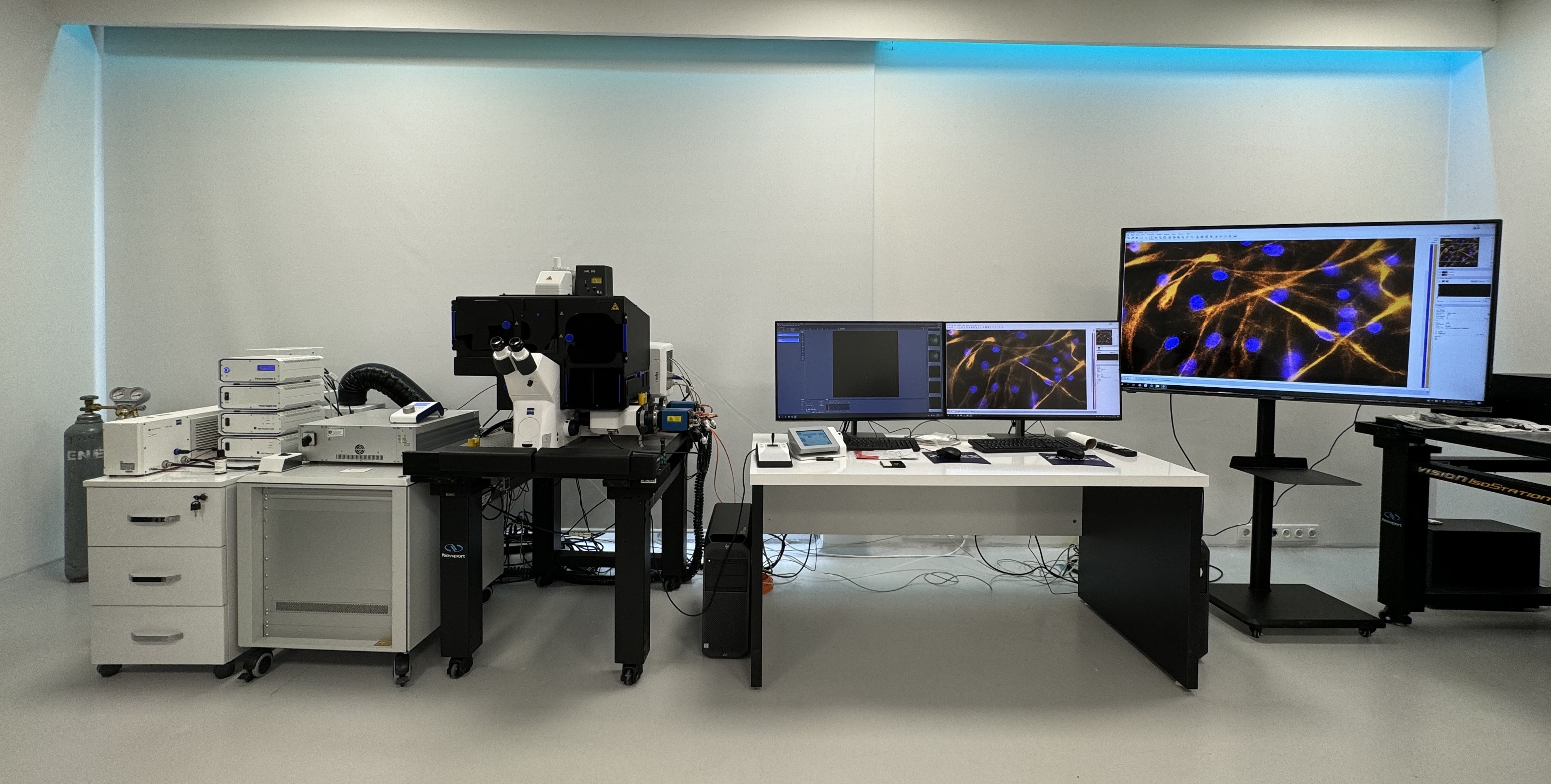
ZEISS Elyra 7 includes a wealth of microscopy techniques to meet your experimental
needs across scales, optimally matching resolution, speed, and sensitivity
requirements to your demanding samples
|
Widefield, DIC |
Widefield (WF) mode (sample illumination with arc lamp), Laser WF mode (sample illumination with laser) |
|
Lattice SIM |
Structured Illumination Microscopy, allowing fast and gentle super-resolution imaging (~120 nm in xy and ~300 nm in z) in 3 dimensions. |
|
Lattice SIM2 |
The new SIM2 module doubles the conventional SIM resolution and achieves up to ~60 nm laterally and ~200 nm axially. The Lattice SIM illumination allows for a better signal to noise ratio, more gentle imaging and can reach up to 255 fps during time lapse acquisition, well suited to capture highly dynamic biological processes in live samples. It further allows deeper imaging of samples up to around 70 µm thickness in comparison to only 20 µm of sample thickness in classical SIM. The speed of this imaging modality is well suited to capturing dynamic biological processes in live samples (significantly faster than regular laser scanning confocal). |
|
Apotome |
Grid-based optical sectioning to create highly contrasted images with high lateral and axial resolution. The Apotome mode is also amenable to SIM2 processing, allowing for high speed acquisition with high contrast, high resolution and low phototoxicity. In combination with SIM2 module, apotome imaging can get lateral rsoltuion down to ~110 nm and axial resolution to ~300 nm. ApoTome imaging is also faster than classic SIM and laser scanning microscopes, allowing for time lapse imaging of faster cellular/tissue dynamics. |
|
SMLM |
Single-molecule localization microscopy like dSTORM, PALM and PAINT allowing resolution down to ~20-30 nm in xy and ~50-80 nm in z. |
|
Dual filter sets for Duolink optimized for dual color and double dual color applications |
Filter sets are optimized for dual camera applications, maximum sensitivity, minimal cross-talk and reduced autofluorescence |
|
Incubation |
Co2 ,Humudity,Temprature |
|
Bio Apps |
|
|
Arivis 4D |
|
|
Available Optics |
|
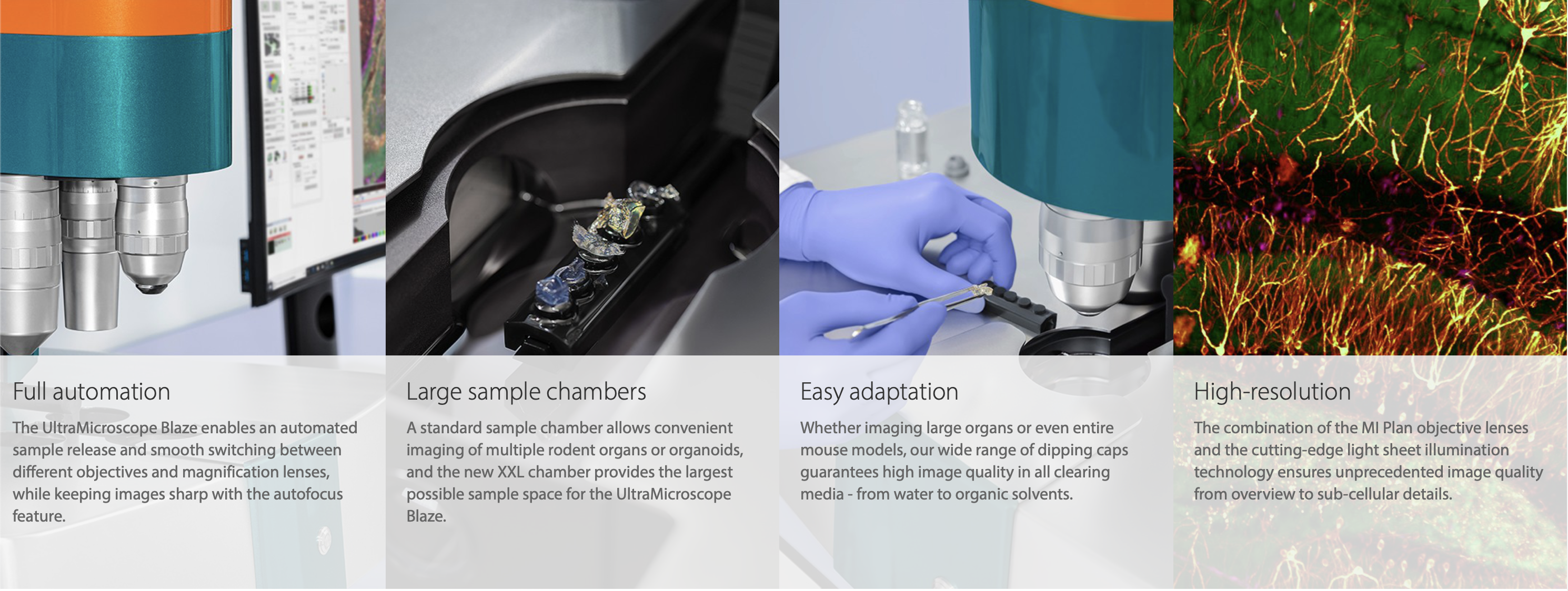
UltraMicroscope Blaze™
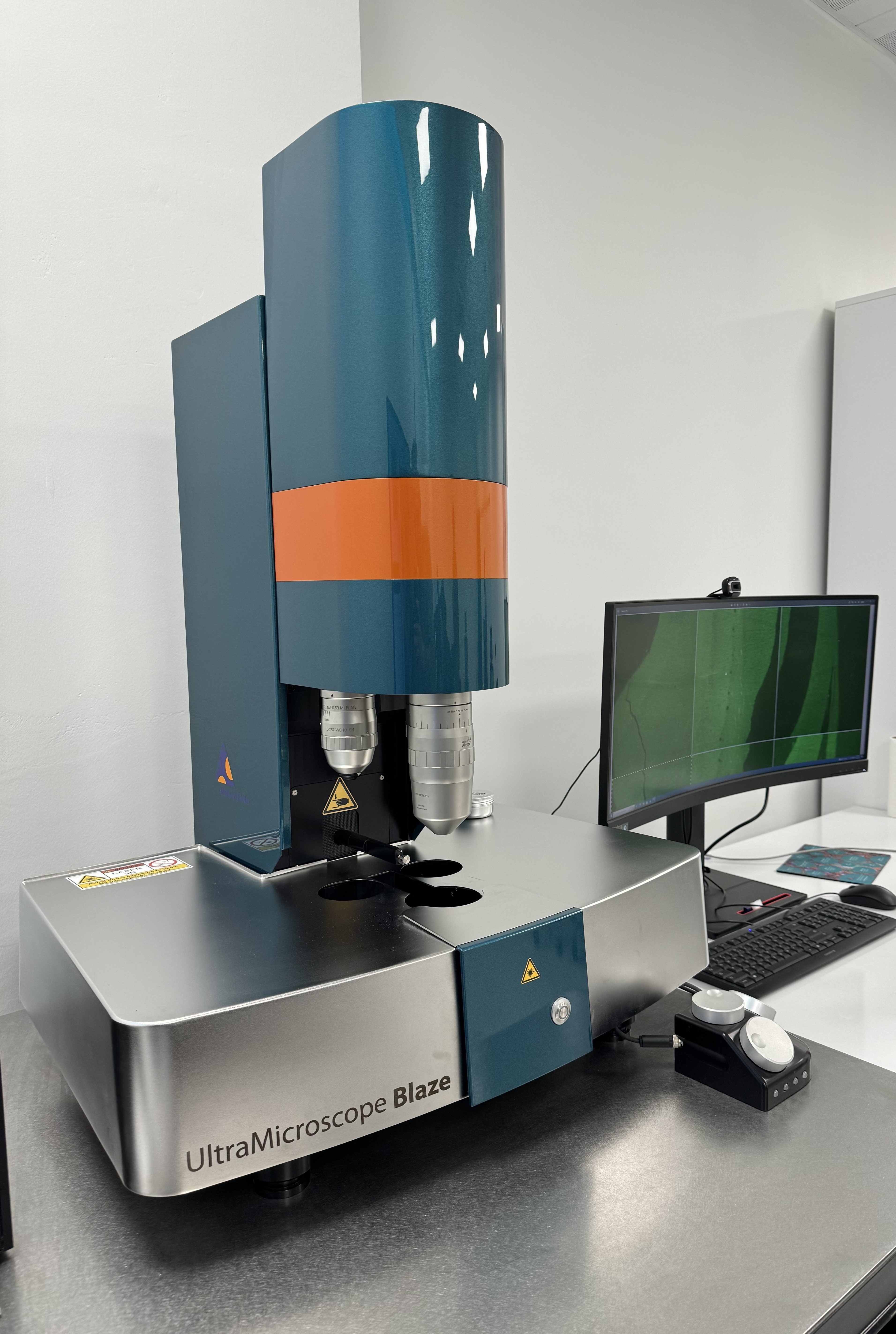
The UltraMicroscope Blaze is the only fully automated light sheet microscope for imaging large or multiple cleared samples at subcellular resolution. Our pioneering UltraMicroscope technology combined with cutting-edge light sheet optics ensures excellent data quality.
A standard sample chamber allows convenient imaging of multiple rodent organs or organoids. Additionally, the new XXL chamber represents a powerful upgrade, specially designed to provide the largest possible sample space for the UltraMicroscope Blaze. It enables fitting of samples as big as a human kidney or whole adult mouse models.
ZEISS Axiovert 5
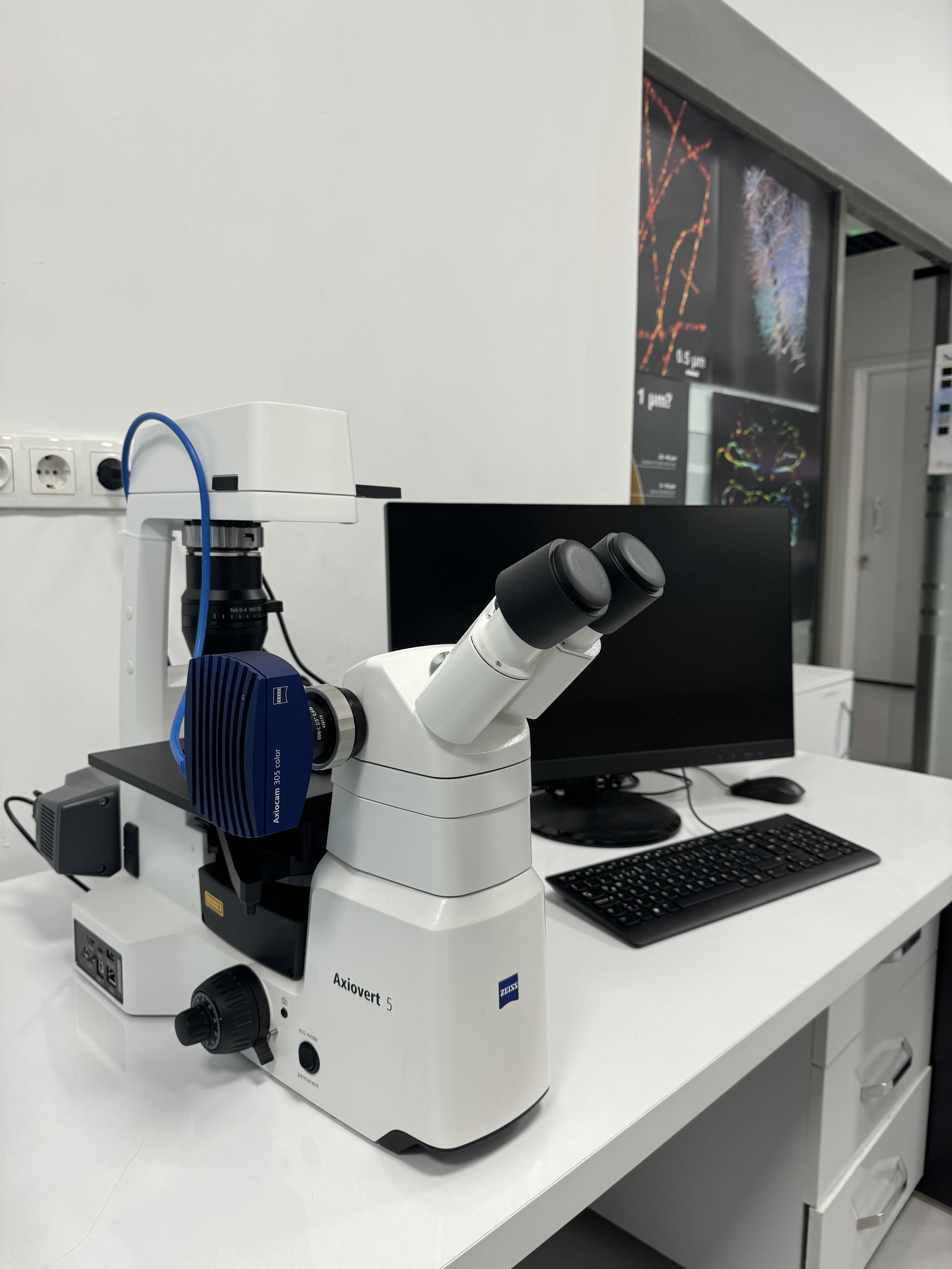
Smart Inverted Microscope for Cell Culture and Research. Integrated Colibri 3 for multichannel fluorescence documentation.
|
Availabe Optic Name |
Magnification |
N.A |
İmmersion |
W.D |
|
Zeiss A-Plan-PH |
5x |
0.15 |
Air |
11.7 |
|
Zeiss A-Plan-PH |
10x |
0.25 |
Air |
8.5 |
|
ZEISS Plan-Neofluar |
20x |
0.50 |
Air |
2.0 |
|
ZEISS Plan-Neofluar |
40x |
0.60 |
Air |
2.9 |
Available Camera:Axiocam 305
Available Excitation Bands:

ZEISS AXİO SCOPE A.1
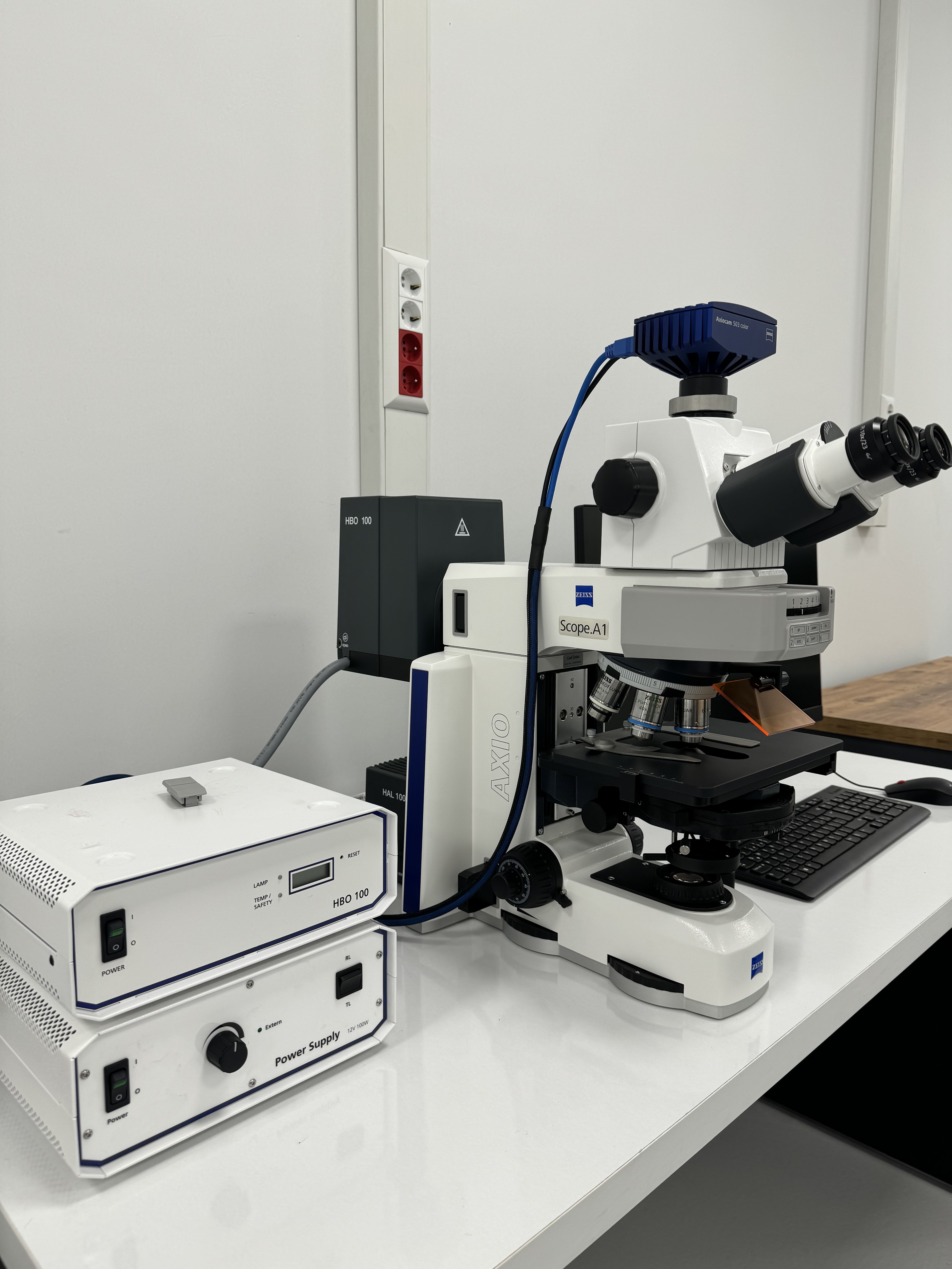
High quality fluorescence , bright field and transmitted polarized documentation with an upright microscope. System has HBO Fluorescence light source.
|
Availabe Optic Name |
Magnification |
N.A |
İmmersion |
W.D |
|
ZEISS Plan-Neofluar |
5X |
0.16 |
Air |
18.5 |
|
ZEISS Plan-Neofluar |
10X |
0.30 |
Air |
5.2 |
|
ZEISS Plan-Neofluar |
20X |
0.50 |
Air |
2 |
|
ZEISS Plan-Neofluar |
40X |
0.75 |
Air |
0.71 |
|
ZEISS Plan-Neofluar |
100X |
1.3 |
Oil |
0.2 |
Available Camera: Axiocam 503 Color
Available Floresans Filters:
|
Excitation |
Beamsplitter |
Emission |
|
G 365 |
FT 395 |
BP 445/50 |
|
BP 470/40 |
FT 495 |
BP 525/50 |
|
BP 546/12 |
FT 560 |
BP 575-640 |
ZEISS STEMI 305
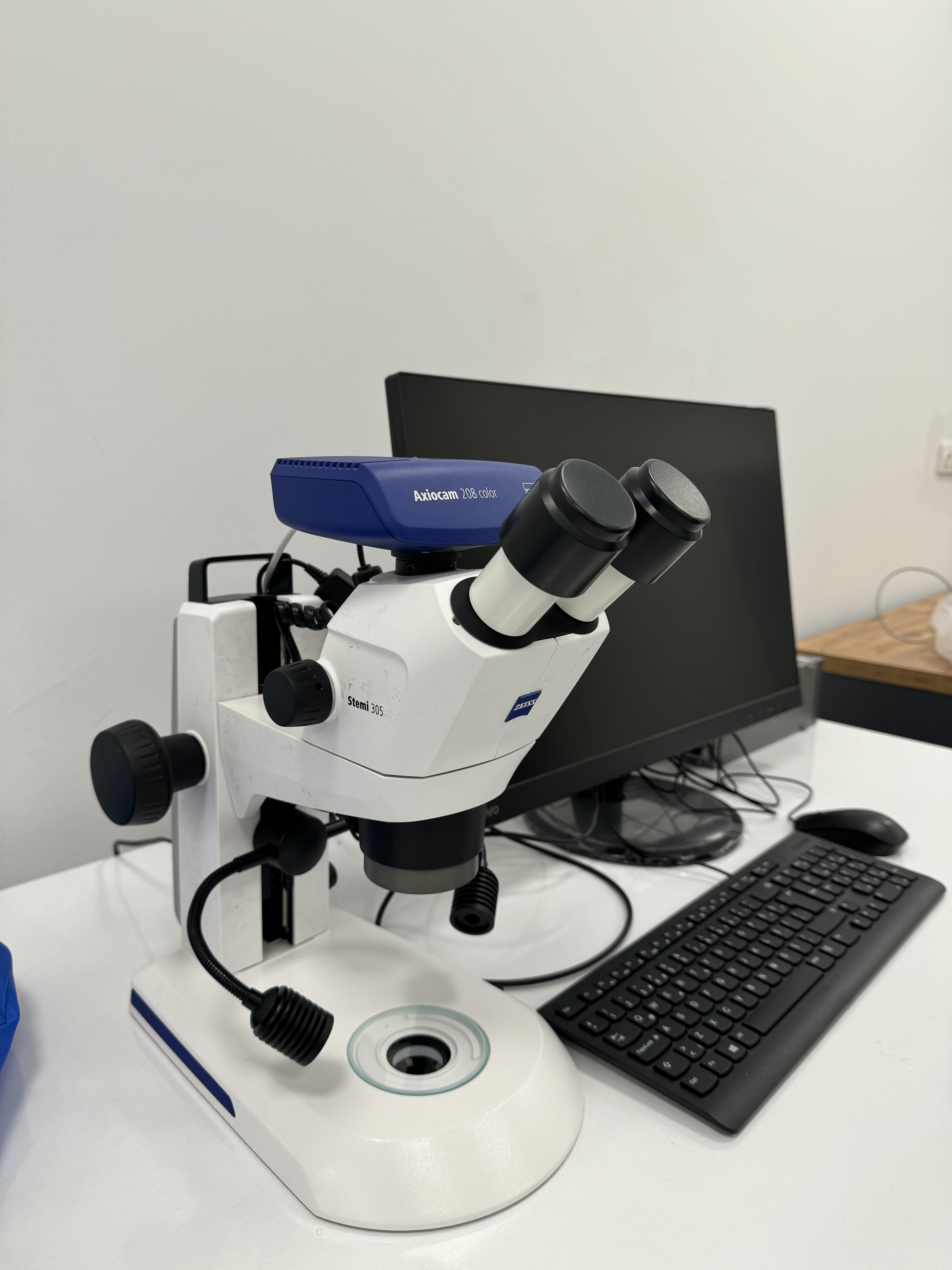
Stemi 305 is compact, easy-to-use stereo microscope with 5:1 zoom. Everything is integrated: long-living LED illumination for reflected and transmitted light and documentation, too.
System has;
- Axiocam 208- 4K camera for documentation.
- 1X objective with 110 mm FWD min zoom 8x ( 28.8 mm object field) max zoom 40x(5.8 mm object field).
- Gooseneck for reflected light
- Brightfield and darkfield for transmitted light
- Maximum Resolution: 200 Lp/mm – 2.5 μm