This study, conducted by NÖROM researchers, reveals that the brain's adaptation mechanism to visual stimuli is impaired in migraine patients.
For migraine patients, complex visual environments—such as supermarket shelves, high-contrast striped patterns, or crowded streets—often turn into a distressing overload. A new study conducted within the Neuroscience and Neurotechnology Center of Excellence (NÖROM) and published in The Journal of Headache and Pain has utilized advanced neuroimaging methods to shed light on why the brains of individuals with migraines struggle more during such visual tasks and why they experience cognitive fatigue more quickly.
The research provides significant findings on how changes in the brain networks of migraine patients without aura affect the ability to filter and organize visual information from the outside world.
The Brain's Ability to Silence "Noise" is Disrupted
In healthy individuals, the brain conserves energy by gradually reducing its response to repetitive stimuli (repetition suppression); this is a fundamental adaptation mechanism that prevents the brain from becoming fatigued by unnecessary information. However, 3T fMRI (functional magnetic resonance imaging) analyses within NÖROM revealed that this mechanism functions differently in migraine patients.
Key Scientific Discoveries:
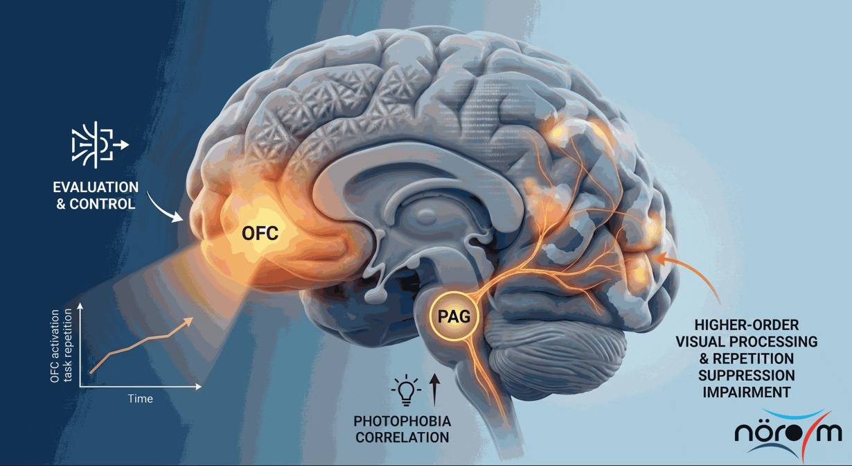
- Loss of Adaptation and Overactivation: During complex visual tasks, the brains of migraine patients operate with much higher energy expenditure in attention and control centers (salience and dorsal attention networks). Particularly in high-level centers that process visual information, the normally expected "sensory habituation" reflex is not observed in individuals with migraines.
- Energy Consumption and Performance Decline: Although individuals with migraines and the healthy control group showed similar success at the beginning of the study, the accuracy rates of migraine patients exhibited a decline toward the end of the task. This situation is considered a concrete indicator of "mental fatigue" resulting from the excessive effort exerted by the brain to manage the visual load.
- Orbitofrontal Cortex and Compensation Mechanism: The orbitofrontal cortex (OFC), the brain's evaluation and control center, becomes more active in migraine patients as the task duration increases. The increase in activity in this region reflects the intense conscious effort patients exert to maintain their performance.
- Light Sensitivity and Pain Centers: It was determined that as sensitivity to light (photophobia) increases, activity changes in the periaqueductal gray (PAG) region, the brain's pain modulation center, also increase. This indicates that visual stimuli are processed in individuals with migraines not as neutral information, but as a "distressing/aversive" experience.
This research, carried out at NÖROM, proves that migraine is not just a headache attack, but also involves structural and functional differences in the brain's capacity to process and regulate sensory information during the periods between attacks. These findings provide a model explaining at a network level why migraine patients avoid visual clutter in daily life and why they experience cognitive fatigue more quickly. In the future, monitoring these specific brain changes may enable the development of next-generation biomarkers in the diagnosis and treatment processes of migraine.
Onlat, Z.C., Ustun, S., Guzel, I. et al. BOLD repetition enhancement in the orbitofrontal cortex during complex visuospatial processing in migraine without aura: a shift in periaqueductal gray - cortical coupling?. J Headache Pain27, 75 (2026). https://doi.org/10.1186/s10194-026-02327-w
Our Center NÖROM Has Joined EBRAINS AISBL as an Associate Member
An Event Titled “From Digital to Traditional, Awakening from Screen to Field” Was Organized
NÖROM Research: Protective Effects of Boric Acid on Sepsis-Induced Organ Damage
Prof. Hayrunnisa Bolay Belen Delivered the Final NÖROnoM Seminar of the Academic Year